-
Category
Craniomaxillofacial Surgery
Orthopedic Surgery
Spine Surgery
Orthopedic Implants
Hip Surgery
Knee Surgery
Pectus Excavatum
Bone Graft
Disinfectants
Healthcare
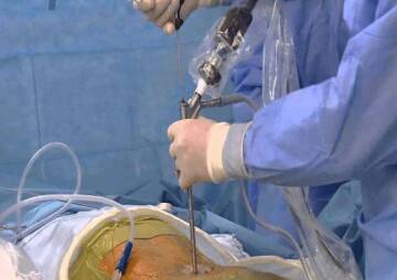
How orthopedic pedicle screws use in spine surgery
Pedicle screws are innovative surgical instruments with the aim to reinforce spinal fusion in the various injury cases of spinal column for several decades.
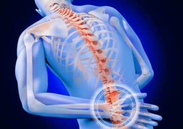
Pedicle screws are innovative surgical instruments with the aim to reinforce spinal fusion in the various injury cases of spinal column for several decades. Spinal fusion is a surgical technique to join one or more vertebrae simultaneously to prevent the motion between them. Curvature abnormality of the spine (scoliosis or kyphosis), spinal vertebrae injury, protrusion of the cushioning disk between vertebrae (slipped disk, herniated nucleus pulposus) and weak or unstable spine caused by infections or tumors are some cases that spinal fusion has to be recommended and appraised.
Pedicle screws for spine and anterior interbody cages, and posterior lumbar cages are three primary methods of spine surgery instrumentation. The pedicle screw is a particular type of bone screw designed for implantation into a vertebral pedicle that provides a means of grasping a spinal segment in any level of spinal column (cervical, thoracic and lumbosacral). In this article the pedicle screw for spinal fusion and the process of preoperative requirements and application of screws and rods are reviewed. At the end, risks and potential complications of the pedicle screws have been discussed briefly.
Pedicle screw for spinal fusion
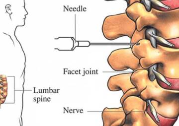
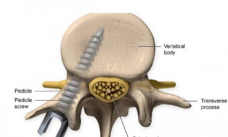
Pedicle screw instrumentation is a technology that has been used to stabilize the thoracolumbar spine and now almost exclusively utilized when securing fusion constructs, due to the intended improved fusion rates and rigidity afforded by these constructs. The pedicle is a dense stem-like structure that protrude from the posterior of a vertebra. There are two pedicles per vertebra that connect to other structures (e.g. lamina, vertebral arch). The pedicle screw system consists of the screws and the rods. The screws themselves do not fixate the spinal segment, but provide immovable anchor points that can then be connected with a rod. The screws are placed at two or three consecutive spine segments (e.g. lumbar segment 4 and 5) and then a short rod is used to connect the screws. This construct can suppress the motion between the segments that are being fused.
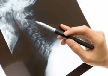
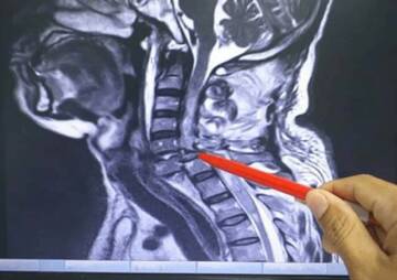
As the pedicles in the lumbar vertebrae are thicker and thus easier to cannulate and generally have protrudes that do not include significant neural or vascular structures, pedicle screws have been applying more often in the lumbar spine. Although pedicle screws are most often used in the lumbar (lumbosacral) spine, they can be implanted in the thoracic and cervical vertebra. To determine the depth and angle for screw implantation, fluoroscopy or conventional x-ray are using by the surgeons. A receiving channel is drilled and the screw is inserted.
 Assistive techniques that can be used are fluoroscopy and stereotactic navigation. Imaging has consequences in increased morbidity associated with radiation exposure, increased time expenditure, and possible workflow interruption, but the increased visualization of a pedicle’s trajectory is beneficial. Several reports have confirmed the high accuracies with each of these three core techniques. However, due to the definition diversities of accuracy and varying radiographic analyses, it is difficult to compare studies to decide which methods are superior.
In some cases there are several difficulties to apply pedicle screws, for instance performing cannulation on the vertebrae within the mid-thoracic spine and vertebrae that have altered morphology due to scoliosis or other deformities. In these circumstances orthopedic spine surgeons should benefit from using aiding technologies.
On the other hand, in the thoracolumbar spine the pedicles should theoretically be fairly cannulated with ease via a free-hand method, given appropriate training and experience. There is, however, a steep learning curve in the technique for placing the pedicle screws, and only surgeons comfortable and experienced with the technique should use them. After all, despite these global recommendations the surgeon’s discretion based on the experience is preeminent to choose the appropriate technique.
Visit Health News Center's pedicle screws: Pedicle Screws List
Pedicle screw insertion
By starting the surgery the proper levels of fixation should be approved as the spine is exposed.
Pitfall: Unstable injuries
In unsteady injuries, a different orientation of the pedicle trajectory might be seen in the segments above and below the level of injury because of the displacement and rotation after the impact. In severely unstable injuries, care must be taken not to cause more dislocation while using the pedicle drill. If it’s necessary, by Image intensifier starting point and trajectory must be confirmed. The pedicle integrity must be assessed for all levels of instrumentation before the operation commencement.
Entry Points
Lumbar spine
The entry point of the pedicle screw is clarified as the confluence of any of the four lines:
- Pars interarticularis
- Mamillary process
- Lateral border of the superior articular facet
- Mid transverse process.

Thoracic spine
The entry point of the pedicle screw for the lower thoracic segments is clarified after determining the intersection of the mid portion of the facet joint and the superior edge of the transverse process. The specific entry point will be just lateral and caudal to this intersection. On the more proximal thoracic levels, the entry point tends to be more cephalad.
Landmarks:
- Lateral border of the superior facet
- Lateral border of the inferior facet
- Ridge of the pars interarticularis
- Transverse process

Opening of the cortex
A burr or rongeur could be used to open the superficial cortex of the entry point.

Cranial-caudal angulation
A pedicle probe is used to guide down the isthmus of the pedicle into the vertebral body. The proper protrusion of the pedicle probe in the cranial caudal direction transpire by intending for the contralateral transverse process thereby intending to be parallel to the superior endplate.

Medio-lateral inclination
Depending on the rotation of the vertebra and the location up to 45° in L5 or 0° in T5, the medio-lateral inclined direction is confirmed. At the depth of insertion, it’s primarily concerned to prevent medial penetration of the spinal canal superficially and lateral or anterior penetration of the vertebral body cortex. Ideally, the two screws should converge but stay entirely within the cortex of the pedicles and body.

Screw insertion
By using a pedicle sounding device the satisfactory completed intraosseous trajectory must be confirmed by pedicle and body palpation after the pedicle track has been fulfilled. In the whole process of the track creation, radiographic approval can be procured.

Note: Depending on the surgeon distinction, a mono- or a polyaxial screw might be selected for the operation.
A pedicle screw of proper diameter and length is carefully inserted and applied into the established trajectory.

Pedicle Screw Risks and Potential Complications
The breakage rate of modern pedicle screws has been reduced to about one in 1,000, while the breakage rate was approximately 10% in the 1980's. A research conducted by the North American Spine Society by 350 physicians on 2,500 patients showed that the complication rate with using pedicle screws in spinal fusion surgery is low. The damage chance of nerve root and the chance of infection, are about one in 1,000, and a 2% to 3% respectively.
The safety and effectiveness of pedicle screws was concerned primarily. However, the initial argument has been promising resolved and for definite situations the pedicle screws are now approved by the FDA for use in the lower (lumbar) spine. The screws and rods may be safely removed with a following back surgery because of the appropriate growing of the bone graft the stability could be obtained properly.
However, the removal is not suggested by most surgeons unless the patient complains discommodity because of the pedicle screws (5% to 10% of cases). In some case reports demonstrated pedicle screws cause harm within the spinal canal or disc space due to the loosening misplacement after surgical correction of adolescent idiopathic scoliosis.
By using the pedicle screws in the posterolateral gutter fusion, the spinal fusion rates have been enhanced from about 60% to 90%. In addition, on the opinion of many surgeons, the pedicle screws improved the recovery of the patients due to the immediate stability for the spine and early ambulation of the patient.
The lower spinal levels are less vulnerable to serious neural damage because of the medially directed screws, as the elements of the cauda equina are much less tending to injury. However, recently a considerable adoption has been obtained for the pedicle screws in the thoracic spine due to the inherent biomechanical improvement, there is a much lower margin of slither confidently, as straying screws are able to injure the spinal cord and other structures intimately related to the vertebrae, including the thoracic pleura, esophagus and intercostal and segmental vessels. After all, other structures within the thoracic cavity comprising the thoracic duct, azygous vein, inferior vena cava, and aorta are vulnerable likewise.
That is the concise of technological innovative remedy platform aiming progress of surgical experiences and improvements in clinical efficiency.
With a considerable potency over the treating multiple spinal pathologies, Health News Center Company offers a prepossessing variety of spinal surgery facilities produced by our country trustworthy manufacturers and exported to several countries all over the world that could be the seemly choice of surgeons to help the patients regain their health.
Refrence: surgeryreference