-
Category
Craniomaxillofacial Surgery
Orthopedic Surgery
Spine Surgery
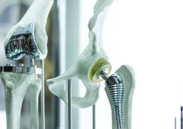
Orthopedic Implants
Hip Surgery
Knee Surgery
Pectus Excavatum
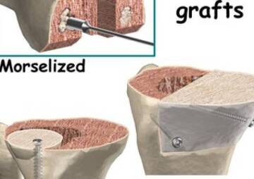
Bone Graft
Disinfectants
Healthcare
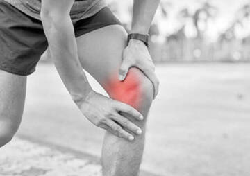
Knee Anatomy And Common Injuries
Knee Anatomy, Injuries, and Appropriate Treatments for Any Injured Part of the Knee are Important Topics to Learn. In this article, everything is written about this topic.
How well do you know about knee anatomy? Why is it important to know a lot about the knee joint anatomy? The knee is one of the most complex joints in the body. Your knee anatomy plays a significant role in knee-injury prevention. If you have a large amount of trauma to the knee, you are more likely to develop injuries to these structures. So, learning about knee anatomy can help you understand whether or not you have some damage to your knee or not. Everything regarding knee anatomy is covered in this post so that you may take better care of your knee with a better understanding.
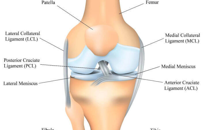
Knee Bones

The bones of the knee are composed of the femur (thigh bone), tibia (shin bone), and patella (kneecap). These bones are kept in place by the knee joint.
An important part of knee joint anatomy is the patella. The patella is the rounded-shaped bone outside the knee that connects the femur to the shinbone (the tibia). The location of the patella can be determined by an examination of the menisci (the inner thigh bones). The location of the patella will also determine the direction of knee movement. When the patella lies close to the shinbone, it is called a flat foot, and if the patella lies between the femur and the knee, it is known as a high-arched foot.
Knee Cartilages

In the knee joint anatomy, two important cartilages are very sensitive to external forces and abrasion:
Menisci
There are two menisci in the knee joint, the medial Meniscus (inside the knee) and the lateral Meniscus (outside the knee). What is the role of these in the knee? The menisci are crescent-shaped discs that act as a cushion and absorb shock between the two bones. This structure causes the knee bones to move in different directions without rubbing against each other. Knowing the meniscus anatomy is also important because most knee problems originate from this part.
Inner knee pain is sometimes caused by a tear and wear of the Medial Meniscus. A meniscus tear can be caused by a direct blow to the Meniscus, such as when your knee hits a hard object while running or landing awkwardly. Alternatively, the Meniscus can be injured through rapid (accelerated) movements like jumping or changing mid-air. In addition, repetitive motions or excessive strength training may also lead to a meniscus tear. Meniscus tears are very painful and need urgent treatment.
Most individuals who have a Meniscus tear choose surgery. It considers as the most effective option since the injured Meniscus can make the leg unstable ( buckle and give way), create pain and inflammation, or even cause the leg to lock up (or become trapped).
Articular cartilage
The articular cartilage in the knee resembles the flexible cartilage that lines chicken bones. This tough, slippery tissue is also similar to the cartilage (mucilage) that exists on other ends of human thigh bones. The cartilages on these ends help the joints absorb shock, provide lubrication, and cushion the joints.
Surgical therapies are generally not recommended for people with Articular Cartilage Lesions in the knee. Before surgery, the patient's medical history is recorded, and a physical examination is undertaken to decide whether surgery is needed in cases of Articular Cartilage Injuries or not. Suppose a patient undergoes surgery without first seeking medical advice. In that case, there are greater chances that the patient may suffer further damages and/or complications after the surgery.
Suppose the condition of Articular Cartilage Injuries is diagnosed as an acute case. In that case, the doctor may recommend the application of a local steroid ointment. This local steroid injection is done to reduce the pain, swelling and promote healing. As soon as the doctor prescribes local steroids, the affected area must be kept clean and dry. Surgical procedures like arthroscopic knee vasectomy, knee joint replacement, and knee cap elevation help reduce the pain and promote healing. However, in some cases, non-surgical treatment can also prove to be effective.
Knee Anatomy Ligaments

A ligament is a strong tissue that connects two bones together, keeping them from moving too much and helping to keep them stable. There are four ligaments in the knee:
Anterior Cruciate Ligament (ACL)
The anterior cruciate ligament is the only one of the four large ligaments located on the outside of the knee that extends over the surface of the lower leg. It acts to stabilize the rotational motion of the lower leg.
Diagnosis of ACL tears, their treatment and prevention: The incidence of ACL injuries or Sprains and strains varies significantly by sex and sport. A qualified surgeon can do an ACL injury diagnostic test and MRI of a knee and forecast the likelihood of injury and recovery time. In many cases, MRI alone cannot correctly determine if an ACL tear is present or not, especially with younger athletes. For this reason, a consultation with an athletic trainer or doctor is highly recommended before treatment and/or diagnosis begins.
Pain, a loud pop or a "popping" sensation in the knee, loss of range of motion, rapid swelling, and instability of the knees are the primary symptoms of an ACL injury. If an ACL tear is suspected, a consultation with a doctor should be made first. Knee braces are available for this purpose and a variety of other treatments, including physical therapy and a type of knee immobilizer to help strengthen the ligaments. Treatment can take a while, depending on the severity of the injury and the extent of the ligament damage.
Posterior Cruciate Ligament (PCL)
The posterior cruciate ligament, also known as the PCL, is the most significant and robust ligament on the lower leg. It has two distinct parts: one that comes from the back of your shin bone (tibia) straight to the top of the foot called the anteromedial ligament. And another smaller part that goes down to the front of your foot is called the peduncle. This ligament is essential for knee movement and is quite strong, especially during intense activities like sprinting or running.
Posterior Cruciate Ligament (PCL) Tears: it can be injured and torn in many ways. Most injuries to this ligament occur when someone is experiencing an unnatural amount of force while jumping or changing direction. Or if the person is doing something wrong when trying to land from a jump. Symptoms of posterior cruciate ligament tears are sharp or dull pain around the back of the knee, instability, tingling or numbness, stiffness, and swelling.
The diagnosis of PCL tears can be problematic in the case of athletes. The PCL injuries from knee injuries are common. Its incidence has been reported to range between 3% and 37% of all patients with acute knee pain.
Most physicians recommend physical therapy for patients who have the diagnosis of PCL tears. Physical therapy treatment may involve stretching exercises to help prevent the tears from occurring, as well as strengthening exercises to help regain strength once the symptoms appear. Anti-inflammatory medication such as a non-steroidal anti-inflammatory drug (NSAID) such as ibuprofen or naproxen sodium is common to alleviate pain. Non-steroidal anti-inflammatory medications have been shown to reduce pain by 40% when taken before physical therapy treatments. This type of treatment has been shown to improve symptoms in most patients but should be avoided in patients who already have a history of recurrent symptoms.
If conservative treatment does not provide relief and the diagnosis of PCL tear is confirmed by an MRI, the treating physician will recommend surgery. Surgical options vary greatly depending on the severity of the tear and the patient's general health, as well as age at the time of surgery
Medial Collateral Ligament (MCL)
It's a large, thick band of tissue that extends from the thighbone (femur) to a point on the shinbone (tibia) about 4 to 6 inches from the knee's inside side.
MCL Injury: A medial collateral ligament tear is a common sports-related injury common to contact sports like football, basketball, and netball. Injuries to the medial collateral ligament occur in various ways ranging from a sprained MCL to a tear from one of the ligaments connecting the kneecap to the thigh. Other injuries to this area include fractures, dislocations, contusions, strains, sprains, tendinitis, sacroiliac pain syndrome, spondylolisthesis, and a variety of ligament tears.
The first step to treating medial collateral ligament tears involves a physical examination to examine the severity of the tears, where they are located, and establish the extent of the injury. The physician may prescribe anti-inflammatory medication if edema is present. An MRI may be ordered to determine the extent of the tear and determine the nature of the tissue around the area. Once a diagnosis has been established, a treatment plan can be developed that will address the patient's specific needs.
Lateral Collateral Ligament (LCL)
The lateral collateral ligament is an important thin band of soft tissue running along the outer edge of the leg. It attaches the thigh bone (femur) to the tibia, the large bone of the leg that goes down the back of the leg and connects with the ankle. As in the case of the medial collateral ligaments, the primary function of this ligament is to maintain the knee stable while walking or running. This stability is important as the risk of injury increases if the leg is unstable and the knee can rock or bend too much.
Lateral Collateral Ligament Injury: A lateral collateral ligament tear in the knee is a common knee injury causing pain, swelling, and severe bruising. Your LCL is an outer band of tissue found on the exterior of your right knee (the opposite side from where your body begins) that connects your upper leg bones to the thigh bone (the front of the thigh bone). This tissue helps maintain and protect your knee joint against internal forces. Your knee needs to be able to move in various directions without being overly stressed or damaged. Injuries to the LCL can cause your knee to become unstable and give way without warning.
The treatment of an LCL injury is typically a course of anti-inflammatory medications followed by physical therapy to work on the inflammation and swelling. The purpose of physical therapy is to relieve your pain and correct any problems with the ligaments. Your treatment may include crutches and/or a brace to hold your leg while standing.
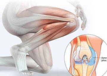
Injuries To The Back Of The Knee
The majority of injuries to the back of the knee involve either one or more of the four large muscles of the quadriceps and hip flexors. These muscles contract to extend the knee. If the muscles become fatigued or overused, they begin to pull the quadriceps and hip flexor tendons into tight spots around the knee. The pulling motion starts at the gluteal muscles, which extend the hip flexor tendons and muscles. It ends with the hamstring muscles, which insert onto the kneecap and pull the hamstring tendon towards the hip bone.
Back of knee anatomy is a very important part of treatment. The tissues and muscles between the bones of the leg help keep the bones in their place and support the weight of the body when it is moving. There are four compartments in the back of the knee. They are the capsule made up of cushions of tissue and the Meniscus, which is the thin line of muscle that runs along the back of the knee. The condyle is the bone on the inner side of the Meniscus, the ischial tuberosity, also known as the isthmic facet, the iliotibial band, and the collateral ligament. This brief description of the back of the knee structure will help the physical therapist or doctor determine the best treatment plan for the patient.
The TakeAway
After reading this article, you will surely realize the importance of the knee structure, including bones, cartilage, ligaments, and the knee tendon anatomy. All of these structures make up the knee joint, and any injury can cause serious problems for you. To take extra care of your knees, you need to be familiar with the anatomy of the knee joint. Doctors also need to know the left knee anatomy and the right knee anatomy to treat knee problems. We've written everything you need to know in this article. Now it's your turn to tell us about your experiences with your knee problems.